Breakthrough Study Links Biomolecular Condensates to Kidney Cancer Development

UTSW Researchers Unveil Key Mechanism Behind Tumor Formation, Paving the Way for Targeted Cancer Therapies
High magnification micrograph of a Xp11.2 translocation renal cell carcinoma (tRCC).
tRCC or translocation renal cell carcinoma is a type of aggressive kidney cancer. About two-thirds of tRCC cases are thought to be caused by fusion of the gene that codes for the transcription factor TFE3. The image is of the tumour stained positively with a TFE3 immunostain.Wikimedia Commons
Published on

A genetic mutation that fuses two genes drives several different cancer types by forming networks of protein interactions that alter gene expression in cells, a study by UT Southwestern Medical Center researchers suggests. The findings, published in Cell, could lead to new treatments for an aggressive kidney cancer and may hold promise for a diverse set of other cancers, the study authors said[1].

"This research identifies a common molecular mechanism shared across diverse cancer-driving oncofusions, revealing a potentially druggable vulnerability."

Dr Benjamin Sabari, Assistant Professor, Cecil H. and Ida Green Center

Dr. Sabari co-led the study with first authors Heankel Lyons, Ph.D., a former member of the Sabari Lab who is now a postdoctoral researcher at Stanford University, and Prashant Pradhan, Ph.D., a postdoctoral researcher at UTSW.

Mutations involving fusions of genes, known as chromosomal translocations, are relatively common in cancers; the different hybrid proteins that they produce, known as oncofusions, are thought to play roles in about 17% of malignancies, Dr. Sabari explained. Why fusing different portions of proteins together causes cancer is understood for a few cancers, but it is unclear for others. These include translocation renal cell carcinoma (tRCC), an aggressive cancer subtype that affects about 4% of adults with kidney cancer and is the most common kidney cancer subtype in children.

About two-thirds of tRCC cases are thought to be caused by fusion of the gene that codes for the transcription factor TFE3 (a protein that’s key for activating specific genes) with one of three other genes: PRCCASPL, or SFPQ. Because the three genes seemingly have nothing in common, Dr. Sabari said, why TFE3 fuses with these specific partners and how this fusion confers malignancy was unknown.

To answer these questions, Drs. Sabari, Lyons, and Pradhan and their colleagues examined tRCC cells from patients treated in UTSW’s Kidney Cancer Program, kept in a biorepository led by study co-author James Brugarolas, M.D., Ph.D., Professor of Internal Medicine and the program’s Director. Using a special stain, they saw that the oncofusion proteins formed biomolecular condensates – dynamic networks of different proteins that segregate themselves within cells – but the “wild-type” (not mutated) TFE3 protein did not. These results suggest that a common feature of the seemingly random fusion partners is the ability to form condensates.

Previous work from the Sabari Lab has shown that condensates can regulate transcription – the process that copies the genetic information in DNA into RNA, an initial step for producing proteins from genes – by selectively capturing key regulatory proteins at target genes. This finding led the researchers to investigate whether oncofusion condensates selectively captured common proteins. When the researchers mixed material extracted from cell nuclei with oncofusion proteins produced by the TFE3 translocation, the proteins readily formed biomolecular condensates, confirming that these hybrid proteins spur condensate formation. Examining proteins enmeshed in the oncofusion condensates identified RNA polymerase II, an enzyme responsible for transcription.

Microscopic image of a purple-colored cell with blue colored organelles.
Biomolecular condensates are specialized droplet-like structures inside cells formed through a process called phase separation. Wikimedia Commons

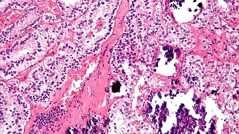
How these three different oncofusions were all able to capture RNA polymerase II was unclear until the researchers compared the amino acid building blocks that make up normal or wild-type TFE3 with those that make up the three mutated oncofusion versions. They found that the mutated versions contained a higher proportion of amino acids that can chemically interact with RNA polymerase II. Upon swapping amino acids between wild-type TFE3 and the TFE3 oncofusion, the oncofusions lost their ability to interact with RNA polymerase II, whereas, conversely, wild-type TFE3 gained the ability to interact with RNA polymerase II, thus confirming the central role of the identified amino acid mixtures in capturing RNA polymerase II and in driving the process of gene transcription.

Cells carrying wild-type TFE3 engineered to carry more of the amino acids from the mutant version adopted cancerous behaviors, becoming more proliferative, invasive, and migratory. The most likely explanation is that RNA polymerase II bound by the mutant amino acids prompted the expression of genes that cause this malignant activity, said Dr. Sabari, also Assistant Professor of Molecular Biology and Obstetrics and Gynecology.

Curious about whether this mechanism might apply to other cancers, the research team combed through a database of known oncofusion mutations found in a variety of cancer types. By examining the amino acids that these mutations code for, the researchers saw combinations similar to those found in mutated TFE3 and found that these other oncofusions also captured RNA polymerase II. These findings suggest that these other oncofusions might be driving diverse cancers through a similar molecular mechanism observed for tRCC.

Finding a way to disrupt these interactions could offer a new way to treat these cancers – a topic that the Sabari Lab plans to pursue in the future.

Reference:

1. https://www.sciencedirect.com/science/article/abs/pii/S0092867425004040

(Newswise/SH)

Related Stories

No stories found.
logo
Medbound
www.medboundtimes.com