Microglia - The Immune Cells Identified as Key Players in Brain Health

Discoveries could inform more targeted therapies for Alzheimer’s and other lipid-rich tissue-related disease
Disease-associated microglia/macrophages are thought to be protective, as they participate in removing lipid-rich waste derived from tissue damage. (Representational Image: Wikimedia Commons)

Using novel genetic and genomic tools, researchers at the Icahn School of Medicine at Mount Sinai have shed light on the role of immune cells called macrophages in lipid-rich tissues like the brain, advancing our understanding of Alzheimer’s and other diseases. The study, published in the March 6 online issue of Nature Communications, represents a step forward in understanding immune cell regulation and its impact on disease progression.

The researchers initially studied genes controlling macrophages, also referred to as microglia when they are in the brain, particularly in response to damage of fatty tissues like the brain. Disease-associated microglia/macrophages are thought to be protective, as they participate in removing lipid-rich waste derived from tissue damage. Therefore, researchers wanted to find factors that promote the “garbage removal” activity of these cells.

Disease-associated microglia/macrophages are thought to be protective, as they participate in removing lipid-rich waste derived from tissue damage. (Representational Image: Wikimedia Commons)
Specialized T cells in the Brain Slow Progression of Alzheimer’s Disease

They identified two influential genes, BHLHE40 and BHLHE41, and used advanced gene editing technology (CRISPR-Cas9) to deactivate them in lab-grown cells. These cells were then transformed into microglia. The resulting microglia lacking BHLHE40 and BHLHE41 resembled disease-associated microglia observed in Alzheimer’s disease, showing improved ability to clear cholesterol-rich waste. Confirmation came from experiments on cultured human peripheral macrophages and microglia from mice lacking these genes.

Through our analysis of single-cell datasets from multiple organs, we've uncovered pivotal regulators of immune cell function essential for tissue health. Our use of advanced models has further validated the critical role played by transcription factors BHLHE40 and BHLHE41, proteins that regulate gene expression by binding to specific DNA sequences, in controlling immune cell responses, presenting potential targets for therapeutic intervention.

Alison M. Goate, DPhil, The Jean C. and James W. Crystal Professor, Chair of Genetics and Genomic Sciences at Icahn Mount Sinai and Senior study author.

Next, the researchers will investigate whether microglia without BHLHE40 and BHLHE41 can help clear away harmful amyloid proteins. In one experiment, the investigators will grow brain cells such as neurons and astrocytes that have harmful Alzheimer’s mutations in a dish and test whether immune cells without BHLHE40 and 41 influence beta-amyloid levels, neurodegeneration, and cytokine response (neuroinflammation). In another, they will inject the immune cells with and without BHLHE40 and 41 into a mouse model of Alzheimer’s to see how they affect the development of Alzheimer 's-like plaques.

The study found that microglia lacking BHLHE40 and BHLHE41, resembling disease-associated microglia in Alzheimer’s disease, showed improved ability to clear cholesterol-rich waste, prompting further investigation into their potential role in clearing harmful amyloid proteins. (Representational Image: Wikimedia Commons)

“We want to see how these cells, particularly those without the two genes, impact Alzheimer 's-related phenotypes in both dish and mouse models. We predict that in mice, microglia without BHLHE40 and 41 will clear away beta-amyloid plaques more effectively than control microglia that have normal levels of BHLHE40 and 41. Also, we're exploring how lack of these genes in the brain immune cells affect other types of cells in the brain such as neurons and astrocytes,” says Dr. Goate.

The paper is titled “BHLHE40/41 regulate microglia and peripheral macrophage responses associated with Alzheimer’s disease and other disorders of lipid-rich tissues.”

The remaining authors of the paper, all with Icahn Mount Sinai except where indicated, are: Anna Podlesny-Drabiniok, PhD; Gloriia Novikova, PhD; Yiyuan Liu, PhD; Josefine Dunst, PhD (Anocca in Sweden); Rose Temizer, PhD candidate (National Institute of Mental Health); Samuele Marro, PhD; Taras Kreslavskiy, PhD (Karolinska Institute); and Edoardo Marcora, PhD.

The study was made possible by funding from various sources, including NIH grants (RF1AG054011, U01AG058635, R56AG081417, U01AG066757, NHLBI R01HL153712, S10OD026880 and S10OD030463) and support from The JPB Foundation and BrightFocus Foundation (A2021014F), the New York State Department of Health stem cell biology fellowship (NYSTEM- C32561GG), America Heart Association (20SFRN35210252), and Graduate Women in Science Fellowship.

(Newswise/NJ)
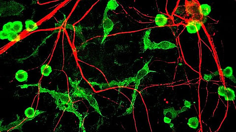
Disease-associated microglia/macrophages are thought to be protective, as they participate in removing lipid-rich waste derived from tissue damage. (Representational Image: Wikimedia Commons)
Researchers Make Precious Headway into a Genetic Form of Alzheimer’s Disease

Related Stories

No stories found.
logo
Medbound
www.medboundtimes.com