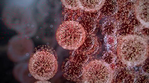
RNA viruses, such as the coronavirus that causes COVID-19, are in a life-and-death race the moment they infect a cell.
These viruses have only minutes to establish their replication machinery inside the host cell before the genetic instructions contained in their vulnerable RNA genomes – which are more fragile than DNA – would otherwise be destroyed by cellular housekeeping. If successful, the virus can go from just a few copies of its RNA genome to a half-million copies incorporated in new infectious particles in less than 12 hours. If not, the virus dies.
In research published online January 24 by the Proceedings of the National Academy of Sciences (PNAS), Morgridge Institute for Research scientists shed new light on these crucial early stages of virus infection and their control. The researchers developed new ways to release viral RNA replication complexes from cells and visualize them in sophisticated ways by cryo-electron microscopy (cryo-EM).
Cryo-EM combines highly advanced imaging with extensive computational analysis to allow scientists to visualize flash-frozen molecules in their native state at molecular to atomic resolution, giving revolutionary insights into biological structure, which can be a powerful foundation for developing therapeutics to thwart disease.
The research team is led led by Paul Ahlquist, director of the Institute’s John and Jeanne Rowe Center for Virology and University of Wisconsin-Madison professor of molecular virology and oncology. The team includes scientists Hong Zhan, Nuruddin Unchwaniwala and Johan den Boon, Morgridge investigator and UW-Madison assistant professor of biochemistry Tim Grant, and co-authors Andrea Rebolledo-Viveros, Janice Pennington, Mark Horswill, Roma Broadberry, and Jonathan Myers.
Most microbe and host genes function in large protein complexes that operate as molecular machines. The structures of these critical assemblies, however, have largely been unknown, greatly limiting understanding and control of the relevant processes. In 2017, using an advanced model virus, the Ahlquist lab provided the first full imaging of a viral RNA replication complex and its striking organization.
They found the parental viral genomic RNA “chromosome” tightly coiled inside a protective membrane vesicle, whose necked channel to the cytoplasm they discovered to be the site of the actual viral RNA replication machinery – the dynamic, multifunctional engine of genome copying - in a previously unknown, 12-fold symmetric, ringed complex that they named the “crown.”
Now, in its new paper in PNAS, the team presents a further leap by revealing the intricate structure of this molecular crown and its component enzyme domains at atomic to near-atomic resolution. These dramatically higher resolution results show how the many distinct functional modules of this replicative engine are arranged, providing an essential basis for working out its assembly, its dynamic operation, and ways to interfere with both.