Getting to the ROOT of your Root Canal Treatment

Did you schedule a root canal appointment at your dentist? Is your tooth pulp throbbing with fear? Yes, you read it right! Tooth has a HEART in the center called PULP.
Root canal treatment also called endodontic therapy is the removal and cleaning of pulp from the pulp chambers and root canals.
Root canal treatment also called endodontic therapy is the removal and cleaning of pulp from the pulp chambers and root canals.Pixabay

Did you schedule a root canal appointment at your dentist? Is your tooth pulp throbbing with fear? Yes, you read it right! Tooth has a HEART in the center called PULP. The RCT or root canal treatment is considered ‘bread and butter amongst dentists! It is the pulp that is keeping your teeth alive and clinics running. Getting away from the lighter side, RCT is the most feared dental treatment among the masses. The fact is, dentists, make the procedure PAINLESS and SAVE TEETH! Root canal treatment also called endodontic therapy is the removal and cleaning of pulp from the root canals. Endodontists specialise in performing root canal treatments.

Root canal treatment also called endodontic therapy is the removal and cleaning of pulp from the chambers and root canals. Endodontists specialise in performing root canal treatments.
Root canal treatment also called endodontic therapy is the removal and cleaning of pulp from the chambers and root canals. Endodontists specialise in performing root canal treatments.Dr. Fatima Hashmi

Why are RCTs needed?

When you get a sweet tooth and eat sugary foods, the food debris remains in the crevices, pits and fissures of teeth. The bacteria (oral flora) in your mouth metabolise sucrose and other carbohydrates to produce acids. These acids dissolve enamel causing cavities/caries. The cavities that do not involve pulp are treated by a routine dental filling. If the holes or cavities are not intervened by the dentist at an early stage these progress into the dentin and then into the pulp tissue or root of the tooth. This leads to the spread of infection in the pulp giving you a toothache. Not all infections result in pain, sometimes a crack in a tooth can open gates for bacteria. PAIN isn’t the only reason you should go to your endodontist!

TELLTALE SIGNS YOU MIGHT NEED RCT

- Toothache.

- Sensitivity to hot and cold foods.

- Lingering pain after eating.

- Swollen gums near the tooth, with pus oozing.

- Discoloration of the tooth.

- Pain on biting.

- Tooth with large cavitation/hole/crack.

A routine dental examination won’t hurt your pockets or teeth. Most of the time symptoms don’t appear unless the infection reaches an advanced stage.

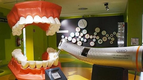
THE TOOTH IS A FASCINATING STRUCTURE OF HARD AND SOFT TISSUES:

The tooth is hollow inside, beneath the hard layers of enamel and dentin with a network of pulp tissue, blood vessels, and nerves. The pulp chamber houses soft pulpal tissue, and blood vessels, which is the source of nourishment for teeth. The floor of the pulp chamber has orifices that lead to root canals.

THIS ARTICLE WILL TAKE YOU INTO THE PULP SPACE-TO THE SOURCE OF PAIN

The tooth crown has a hard layer of ENAMEL which is harder than a diamond. Enamel is made up of mineral hydroxyapatite crystals, and calcium phosphate with no water. It is ironic how diamond-hard enamel is still invaded by bacteria. Dentin lies underneath the enamel. It is mineralized tissue with water content. In the core of teeth lies pulp tracing down along the length of roots.

WHY PULP IN THE TOOTH IS- A PAIN IN THE APEX!

NEUROPHYSIOLOGY OF PAIN

The pulp tissue is highly innervated and vascularised.

It is enclosed in a chamber which leaves no space for the release of pressure resulting in pain. The increased tooth pressure acts directly on sensory nerve receptors. During infection, the pulp tissue gets inflamed and compressed under the hard layers of enamel and dentin and stimulates free nerve fibres (nociceptors). The blood flow in the pulp is more than the surrounding areas i.e. 20-60 mL/min/100g pulp tissue. The pulp tissue lacks alternate/collateral blood circulation thus restricting sufficient blood flow to the site of infection.

LEFT IMAGE-The pulp tissue comes out in the flutes of files. 
RIGHT IMAGE- The intra-oral x-ray help dentist visualise the surrounding bone and other periodontal structures.
LEFT IMAGE-The pulp tissue comes out in the flutes of files. RIGHT IMAGE- The intra-oral x-ray help dentist visualise the surrounding bone and other periodontal structures.Dr. Varun Mhatre

THE ROOT CANAL PROCEDURE/STEPS:

The dentist will first shoot an intra-oral X-ray and load the syringe with local anesthesia. Is this invoking fear in you? Fear not!

The syringe has local anesthesia to make the procedure painless. After a shot of anesthesia, you will feel numb. It is the long needle, the voice of the drill, dental armamentarium that makes you want to come off the dental chair.

1. INTRAORAL X-RAY

The intra-oral x-ray help dentist visualise the surrounding bone and other periodontal structures. The tooth health is evident on x-rays.

RCT is a standard and effective procedure for removing the infected pulp within a tooth. If the infection of tooth is untreated, it can spread to surrounding tissues, resulting in severe bone loss, which aids in further spread leading to space infection or cellulitis.
Dr. Chintan Shetty, BDS

2. LOCAL ANESTHESIA

The dentist will inject local anesthesia around the premises of the treatable tooth. It feels like a bee sting/needle prick.

3. THE DRILL

The dentist then uses an air rotor (2,00,000 R.P.M.) to drill the crown (enamel, dentin) of the tooth to reach inside the pulp chamber to remove dead pulp tissue.

The Lilliputian files come in sizes and are used successively in increasing/decreasing order to give root canals a tapering shape inside.
The Lilliputian files come in sizes and are used successively in increasing/decreasing order to give root canals a tapering shape inside.Unsplash

4. FILING

A dentist uses miniature hand/ air rotor files that fit your itsy-bitsy root canals. These files are given motions of reaming, watch-winding, and push-pull to clean the root canals. This removes debris, dead pulp tissues, and bacteria. The pulp tissue comes out in the flutes of files. It is crucial to remove tissue to avoid re-infection, which would result in the need for re-RCT or extraction.

5. CLEANING/ SHAPING THE CANALS

The Lilliputian files come in sizes and are used successively in increasing/decreasing order to give root canals a tapering shape inside. Along with debris, the filing also creates space for the placement of intracanal medicaments, disinfectants, and sealers.

After each insertion and removal, the canals are cleaned with disinfectants like normal saline (0.9% w/v), sodium hypochlorite, or chlorhexidine, etc. These irrigants will SET YOUR TEETH ON EDGE. These chemicals sterilize root canals, have broad-spectrum antibacterial properties, and inactivate endotoxins.

Gutta-percha is a thermoplastic permanent filling material placed inside canals previously occupied by pulp tissue.
Gutta-percha is a thermoplastic permanent filling material placed inside canals previously occupied by pulp tissue.Dr. Fatima Hashmi

6. CANAL FILLING/OBTURATION

The canals are thoroughly cleaned and dried using absorbent paper points. Gutta-percha is a thermoplastic permanent filling material placed inside canals previously occupied by pulp tissue. The GP cone is radiopaque and hence visible on x-rays. To seal everything from bacterial invasion dentist tops it with a temporary filling material.

Gutta-percha is a thermoplastic permanent filling material placed inside canals previously occupied by pulp tissue. The GP cone is radiopaque and hence visible on x-rays.
Gutta-percha is a thermoplastic permanent filling material placed inside canals previously occupied by pulp tissue. The GP cone is radiopaque and hence visible on x-rays.Dr. Varun Mhatre

7. CROWN/CAP YOUR TOOTH

The crown structure of the tooth has been drilled by the dentist and should be capped to avoid future bacterial invasion.

THIS IS VERY IMPORTANT TO NOTE- Since the nerves and blood vessels along with pulp tissue are removed during the root canal procedure, the tooth becomes brittle hence crown is the pinnacle of the root canal treatment.

Root canal treatment also called endodontic therapy is the removal and cleaning of pulp from the pulp chambers and root canals.
Braces Straighten Teeth is a Half Truth: Embrace those Retainers

Dentists may prescribe you some antibiotics, and painkillers to clear any infection after treatment. Antibiotic course should be completed.

According to studies, a root canal-treated tooth can survive for 10 years or longer.

There is no replacement for natural teeth, and RCT is a repair treatment for your teeth.

How about a trip to the dental clinic?

References:

  1. Dammaschke T, Steven D, Kaup M, Ott KH. Long-term survival of root-canal-treated teeth: a retrospective study over 10 years. J Endod. 2003 Oct;29(10):638-43. doi: 10.1097/00004770-200310000-00006. PMID: 14606785.

  2. Chun K, Choi H, Lee J. Comparison of mechanical property and role between enamel and dentin in the human teeth. J Dent Biomech. 2014 Feb 6;5:1758736014520809. doi: 10.1177/1758736014520809. PMID: 24550998; PMCID: PMC3924884.

Join MedBound - an ecosystem for students and professionals of medical and health sciences.

Related Stories

No stories found.
logo
Medbound
www.medboundtimes.com