The processes by which nerve cells in the brain communicate and how T cells neutralize cancer cells are not fully understood at the cellular level. However, a team of researchers from the Technical University of Munich (TUM) has developed unique reporter proteins that could shed light on these mechanisms.
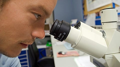
Using an electron microscope, scientists can observe cellular structures at an incredibly detailed level, with resolution reaching sub-nanometer ranges. Even tiny components like mitochondria and nerve cell connections can be seen. However, despite this powerful tool, many important structures and processes are still beyond our ability to observe. According to Gil Gregor Westmeyer, Professor of Neurobiological Engineering at TUM and Director of the Institute for Synthetic Biomedicine at Helmholtz Munich, this limitation is similar to looking at a map of a city, which can provide a general overview but cannot reveal details such as traffic flow and construction projects.
Understanding cellular processes is crucial for intervening in faulty processes or recreating them in artificial tissues and organs. To help in this endeavor, Westmeyer and his team have developed a genetic reporter system that can provide a closer look at processes within and between cells. This system involves protein capsules, which are small enough to be observed with an electron microscope and act as genetic reporters to aid in reconnaissance work within cells.
Identification by barcodes
The protein capsules used in the genetic reporter system are created by the cells themselves, with their genetic blueprints attached to specific target genes. When the target genes become active, the reporter proteins are produced. This approach is based on a standard procedure in light microscopy that uses fluorescent proteins. However, using fluorescent proteins is not suitable for electron microscopy because electron densities, rather than colors, are used to distinguish between different shapes.
To address this challenge, the researchers incorporated metal-binding proteins into capsules of various sizes. These specially designed capsules, known as "EMcapsulins," appear as concentric circles of varying sizes when viewed under an electron microscope. With the help of artificial intelligence, the EMcapsulins can be quickly identified and assigned, much like barcodes.
Making invisible structures visible
The researchers can use the genetic reporter proteins in two ways: firstly, to indicate the activity of certain genes, and secondly, to reveal structures that would otherwise not be visible under an electron microscope. For example, the reporter proteins can help identify electrical synapses between nerve cells or receptors that affect the interaction between cancer cells and T cells. This information can be used to better understand cellular processes and develop new treatments for various diseases.
Felix Sigmund, the first author of the study, explains that by giving the EMcapsulins fluorescent properties, researchers can initially examine structures in living tissue using light microscopy. This approach can reveal striking dynamics and structures that can then be more closely observed using electron microscopy.
Westmeyer suggests that in the future, the reporter proteins could be used as sensors that change their structure when a cell becomes active, providing insight into the relationship between cell function and structure. This information could improve our understanding of disease processes and aid in the development of therapeutic cells and tissues.
To this end, the researchers will also use the new Electron Microscopy Facility at TUM and collaborate with the new TUM Center for Organoid Systems. (PB/Newswise)


