- Home
- About Us
- Contact Us
- Biography
- Column
- MedBound Hub
- InternshipInternship
- Daily Pulse
- Medicine
- Physical Therapy
- InterviewInterview
- Pharmacy
- Biotechnology
- College/InstituteCollege/Institute
- Dentistry
- Blog
- Wellness & Nutrition
- Nursing
- Opinion

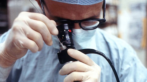
A research team from the lab of Quing Zhu, the Edwin H. Murty Professor of Engineering in the Department of Biomedical Engineering at the McKelvey School of Engineering at Washington University in St. Louis, has combined optical coherence tomography (OCT) and machine learning to develop a colorectal cancer imaging tool that may one day improve the traditional endoscopy currently used by doctors.
The results were published in the June issue of the Journal of Biophotonics, with an image featured on the inside cover.
Screening for colon cancer now relies on human visual inspection of tissue during a colonoscopy procedure. This technique, however, does not detect and diagnose subsurface lesions.
An endoscopy OCT essentially shines a light in the colon to help a clinician see deeper to visualize and diagnose abnormalities. By collaborating with physicians at Washington University School of Medicine and with Chao Zhou, associate professor of biomedical engineering, the team developed a small OCT catheter, which uses a longer wavelength of light, to penetrate 1-2 mm into the tissue samples.
Hongbo Luo, a PhD student in Zhu’s lab, led the work.
The technique provided more information about an abnormality than surface-level, white-light images currently used by physicians. Shuying Li, a biomedical engineering PhD student, used the imaging data to train a machine learning algorithm to differentiate between “normal” and “cancerous” tissue. The combined system allowed them to detect and classify cancerous tissue samples with a 93% diagnostic accuracy.
Zhu also is a professor of radiology at the School of Medicine. Her team worked with Vladimir Kushnir and Vladimir Lamm at the School of Medicine, Zhu’s team of PhD students, including Tiger Nie, started a trial in patients in July 2022.
The study was partially funded by the National Institutes of Health (R01 R01CA237664) and the Siteman Cancer Center (Pre-R01). (SP/Newswise)