- Home
- About Us
- Contact Us
- Biography
- Column
- MedBound Hub
- InternshipInternship
- Daily Pulse
- Medicine
- Physical Therapy
- InterviewInterview
- Pharmacy
- Biotechnology
- College/InstituteCollege/Institute
- Dentistry
- Blog
- Wellness & Nutrition
- Nursing
- Opinion

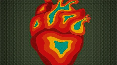
Though neurology and cardiology are separate specialties, the brain and heart have many things in common: specialized cells, electrical signaling, mechanisms of self-regulation, and high energy demands. There’s also a strong relationship between neurologic function and cardiac function, with research finding many links between the two.
Relationships between epilepsy and heart conditions are becoming more and more clear.
People with epilepsy are at increased risk for heart disease, hypertension, atrial fibrillation, and hyperlipidemia. Conversely, people with cardiovascular disease and risk factors are at an increased risk for epilepsy—and the risk goes beyond stroke. Data from the Framingham Heart Study[1] show that hypertension is a risk factor for late-onset epilepsy, even after controlling for stroke.
People with certain types of congenital heart disease also are at increased risk for epilepsy. In some cases, the cardiac and neurological consequences are linked genetically: for example, certain genetic mutations cause channelopathies that can affect both brain and heart.
Over time, seizures can damage the heart’s structure and function, increasing risk of cardiovascular problems, including sudden cardiac death (SCD); this is the concept of the epileptic heart, suggested in a 2020 publication by Richard Verrier, Trudy Pang, and colleagues.
And compared with people without epilepsy, cardiovascular health in people with epilepsy may be fundamentally different, and these differences may affect mortality risk, including risk from sudden unexpected death in epilepsy (SUDEP), according to research by Guilherme Fialho and Katia Lin.
The most common cause of sudden cardiac death (SCD) in the general population is ventricular fibrillation leading to cardiac arrest. People with epilepsy have close to a threefold risk for SCD, compared with the general population.
While there may be some overlap between SCD and SUDEP, “Sudden cardiac death is a result of heart disease, while SUDEP excludes patients with any kind of heart disease that the pathologist can identify,” said Pang. “The SCD group tends to be older, 40 to 75, while patients in the SUDEP group are younger, 20 to 40.”
Pang, Verrier, and colleagues have proposed that chronic seizures—as well as antiseizure medications—damage the heart and coronary vasculature, leading to electrical and mechanical dysfunction. The “epileptic heart” is damaged from repeated hypoxemia, the toxic effects of catecholamines (such as adrenaline) released during seizures, and accelerated atherosclerosis, which can be partially associated with the use of sodium-channel-blocking antiseizure medications.
In their 2020 publication, Verrier, Pang, and colleagues suggest that the increased risk of SCD in people with epilepsy stems from this damage. Preclinical and post-mortem studies have identified cardiac structural changes in people with epilepsy, including myocardial fibrosis and myofibrillar degeneration.
Seizures, particularly generalized tonic-clonic (GTC) seizures, place intense energy demands on the cardiovascular system. During a seizure, the oxygen supply to the heart is reduced; at the same time, the heart is exposed to catecholamines, which “can actually stun the heart,” said Verrier. “When you measure ejection fraction, the percent of blood ejected with each beat, it can be reduced from a normal level of 60% down to 40%.”
Catecholamine levels remain high in the post-ictal period. A study of 30 patients receiving simultaneous video EEG and electrocardiography (EKG) found that blood markers of cardiac stress were elevated in 25% of patients who had GTC seizures, without apparent clinical symptoms.
Animal studies also suggest that seizures alter cardiac ion channel expression over time, leading to increased sympathetic tone and potentially predisposing to arrhythmias. Animal models have shown changes in sodium and potassium channels, sodium-calcium exchangers, and hyperpolarization-activated cyclic nucleotide-gated channels.
In addition to a clinical history, assessing cardiovascular risk in people with epilepsy can begin with a routine EKG. For longer recording times, a Holter monitor or wireless ambulatory patch monitor can collect data over several days to weeks.
Verrier cited several EKG measures that can help to determine cardiovascular risk in people with epilepsy:
P wave irregularities. The P wave represents electrical activation and contraction of the atria. Up to 10% of people with epilepsy are prone to atrial fibrillation, said Verrier. “Any irregularities in the P wave, such as duration or morphology, are worth inspecting in the 12-lead EKG,” he said.
The presence of pathological Q waves, which may reveal a prior silent myocardial infarction. Small Q waves are normal and indicate the depolarization of the muscular wall between the ventricles. Abnormally deep or wide Q waves suggest myocardial damage. “There’s a nearly fivefold increase in myocardial infarction in patients with chronic epilepsy, so inspecting the electrocardiogram for Q waves is quite important,” Verrier said.
ST segment changes. Up to 40% of seizures have been associated with changes in the ST segment, said Verrier. This segment represents the phase between ventricular contraction and ventricular repolarization.
Prolonged QT interval. The QT interval represents the time it takes ventricles to contract and repolarize. “Approximately one-third of patients with drug-resistant epilepsy have prolongation in the QT interval,” said Verrier. “This reflects an increased susceptibility to ventricular arrhythmias, which could also be worsened by certain medications.”
In addition to an EKG, echocardiography can identify myocardial stiffness, which can increase risk for ventricular arrhythmias as well as changes to the left atrium that could predispose to atrial fibrillation.
“I think echocardiography is a valuable tool that should be increasingly used in patients with chronic epilepsies,” said Verrier.
To assess risk for SCD, Verrier and colleagues developed the microvolt T-wave alternans parameter. T-wave alternans is a beat-to-beat variation in the amplitude or shape of the T wave on an EKG. In people with stable cardiovascular disease, the alternans is below 47 microvolts (μv). In people with cardiomyopathies or myocardial infarction, the alternans exceeds 47 mv.
“Nearly 10 years ago, we found that in patients with drug-resistant epilepsy the alternans was in excess of 60 microvolts, well beyond the cut point of risk of life-threatening arrhythmias,” Verrier said. “This was a surprise to us.” Other studies have found that people with drug-resistant epilepsy have T-wave alternans measurements higher than 47 μv, while people with controlled seizures have measurements below this cutoff.
In 2022[2], Verrier and colleagues published a study of 18 people with epilepsy admitted to an epilepsy monitoring unit who were given wireless EKG patch monitors. The T-wave alternans parameter exceeded 60 μv in people having focal to bilateral tonic-clonic seizures, but not those having focal seizures or non-epileptic seizures. The authors suggest that post-seizure mortality in some cases may be due to such cardiac instability.
Incorporating the concept of the epileptic heart into epilepsy care may help identify people at risk, said Pang.
“First and foremost is having a diagnosis of epilepsy, particularly refractory epilepsy,” she said. “And beyond asking about seizure frequency, we also want to think about various cardiac risk factors. Features or clinical signs of potential cardiac injury and the risk for arrhythmia, whether that is by a 12-lead standard EKG or a multi-day ambulatory monitor.”
In addition to tests via EKG and echocardiography, Pang suggested assessing altered autonomic tone using heart-rate variability, as well as assessing classic cardiovascular risk factors, including lipid profiles. “People with epilepsy are at risk for accelerated atherosclerosis if their seizures are poorly controlled,” she said.
In fact, research on the cardiovascular profiles of people with epilepsy has shown that atherosclerosis in people with epilepsy increases the heart’s rate of aging by 10 to 20 years, said Verrier. “So someone who is 40 years old who has chronic epilepsy, their heart may be that of an over 50-year-old,” he said.
Pang said that people with epilepsy who have multiple risk factors for an “epileptic heart” may need regular routine EEGs with ambulatory EKG monitoring to identify arrhythmias, as well as structural imaging to identify structural changes or dysfunction.
“I would go further and say that patients in the highest risk category, for example, those with uncontrolled generalized tonic-clonic seizures and multiple cardiovascular risk factors, those patients should be referred to cardiology for a detailed evaluation,” she said.
Cardiologist Fialho and neurologist Lin are colleagues at the Federal University of Santa Catarina in Florianópolis, Brazil. With others, they have published several studies using EKG, echocardiography, and treadmill testing to understand cardiac differences in people with temporal lobe epilepsy (TLE). The studies have found that lower cardiovascular fitness and increased cardiac stiffness compared with controls, despite similar cardiovascular risk factors between groups.
Cardiovascular fitness is measured by “metabolic equivalent of task,” or MET. It’s a sort of energy expenditure index; one MET is defined as expending 1 kcal/kg/hour, roughly equivalent to sitting quietly. A slow walk clocks in at about 2 METs, while jumping rope is 9 to 10 METs. In treadmill testing, the fittest people have the highest MET scores—and the lowest mortality risk. In fact, research has shown that cardiovascular fitness measured by METs is a more powerful predictor of mortality than other cardiovascular risk factors.
In a 2017 study[3]using treadmill testing, Lin and Fialho found that compared with controls, participants with TLE had lower peak heart rates, exercised for less time, and experienced chronotropic incompetence: The heart was less able to adjust its rate in response to exercise. People with TLE also had lower MET scores: an average of 1.7 METs lower than the control group.
“MET is a very strong marker of risk for not only cardiovascular death, but all-cause death,” said Fialho. He cited a landmark 2002 study that found that every 1-MET increase in fitness was linked with a 12% decrease in mortality risk. Other studies, he said, have found higher numbers: 15%, 18%, even 20% fewer deaths with every 1-MET increase.
“So this 1.7 METs less measure was a very big deal,” he said. “If you put those individuals [with epilepsy] into our classic Framingham risk table[4] or use other scores to study their cardiovascular risk, they would be considered low risk. But they are not low risk.”
The people in Fialho’s studies had an average age of 37 and had been living with TLE for an average of 22 years. While they may have been less fit because they had been discouraged from exercising or because of other lifestyle factors, the increased cardiac stiffness reduces the heart’s ability to pump blood and distribute oxygen, which directly impacts fitness.
Statistical analysis showed that 52% of the cardiac stiffness could be explained by autonomic dysfunction, with another 6% linked to carbamazepine therapy and 6% to polytherapy.
“We need trials, and until we have those, we can’t really say [what is going on],” Fialho said. “But we have to remember that people with epilepsy may be at increased risk of arrhythmia and sudden death.”
References
[1] https://onlinelibrary.wiley.com/doi/epdf/10.1111/epi.17108
[2] https://www.epilepsybehavior.com/article/S1525-5050(22)00330-4/abstract
[3] https://www.sciencedirect.com/science/article/abs/pii/S0920121117300943?via%3Dihub
[4] https://www.framinghamheartstudy.org/fhs-risk-functions/cardiovascular-disease-10-year-risk/
(Newswise/MKB)