What Happens When our Heart Experiences Blood Flow Reflux?

Aortic regurgitation results from the blood from the aorta flowing back into the left ventricle during the diastolic phase of the heart's activity.
Aortic regurgitation results from the blood from the aorta flowing back into the left ventricle during the diastolic phase of the heart's activity (Pixabay)

The heart is an exquisitely developed organ that primarily has four chambers: the right and left atria, and the right and left ventricles. For these chambers, the four valves serve as a bypass.

The right atria to ventricle flow are regulated by the tricuspid valve.

The left atria to ventricle flow are controlled by the mitral valve.

The aortic valve regulates the flow from the left ventricle to the aorta which is the main blood vessel.

Right ventricle to pulmonary artery flow is controlled by the pulmonary valve.

Pathological issues with these leads, particularly stenosis and regurgitation. The focus of this article is aortic regurgitation.

Regurgitation: What is it?

The valves' primary function is to stop blood from flowing backward. However, the valves don't close because of an underlying pathological issue. Aortic regurgitation results from the blood from the aorta flowing back into the left ventricle during the diastolic phase of the heart's activity.

Heart in the throat!!

 What does it represent?

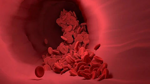
Patients with aortic regurgitation can see a palpable pulse of an aneurysm on the aortic arch. Aortic insufficiency results from the aortic valve's malfunctioning in aortic regurgitation, which causes the blood to flow back into the left ventricle. This causes the blood flow from the aorta to diminish, which causes the carotid artery to collapse. This condition is known as the CORRIGAN SIGN, and it is brilliantly illustrated in the video up top. It is among the crucial signs of aortic regurgitation.

The story of this sign begins with Sir Dominic Corrigan, the physicist who first noticed it. Thomas Watson later compared it to a water-hammer toy in 1844, 11 years after the discovery, and as a result, the name Corrigan sign was replaced with Watson pulse. Collapsing pulse, water hammer pulse, and pulsus celer are some of the alternate names.

What makes it hazardous?

As is well known, the left ventricles pump oxygenated blood throughout the body, and a reduction in blood flow to the aorta affects the delivery of blood to other organs. As blood flows back into the left ventricle, the left ventricle's pressure increases and the stroke volume reduces as a result. Low cardiac output can result in hypotension and, in extreme circumstances, cardiogenic shock.

What causes it?

Rheumatic disorders, where antibodies target our body cells, in this case, the valve, are the most frequent cause. Congenital abnormalities, the presence of only two leaflets (in this situation, the tricuspid valve's one leaflet is attached to the aortic valve), an aortic aneurysm, and degenerative illnesses are other common causes. The common risk factor is high blood pressure. Blood flow increases during this, which causes the valve to dilate and eventually become difficult to seal properly, resulting in regurgitation. Another cause could be connective tissue disorders and aortic inflammation.

Aortic regurgitation results from the blood from the aorta flowing back into the left ventricle during the diastolic phase of the heart's activity (Pixabay)
5 Foods for your Joint Health

How do patients with aortic regurgitation appear?

Clinically, there are two types of aortic regurgitation: acute and chronic. Acute aortic regurgitation causes patients to experience extreme breathlessness because less blood is being produced and less of it is getting to their organs, which means that they are receiving less oxygen.

• Heart failure

• Heartbeat speeding up as pressure builds

Cyanosis and pulmonary edema

Palpitations, a feeling of a pounding heartbeat, shortness of breath, chest pain, and sudden cardiac death are all symptoms of chronic aortic regurgitation.

Aortic regurgitation results from the blood from the aorta flowing back into the left ventricle during the diastolic phase of the heart's activity (Pixabay)
World Mental Health Day: Correlation between Mental Health and Gut

There are a few key indicators that Aortic regurgitation is present.

1. The De Musset sign, which displays head nodding for each heartbeat

2. When the popliteal systolic pressure is higher than the brachial systolic pressure, this is known as the "Hill sign."

3. Corrigan pulse, also known as a "water hammer pulse," is characterized by excessive arterial pulsations that are visible and cause a strong and rapid collapse in pulse as demonstrated in the video.

4. Quincke sign: Capillary pulsations are felt when pressure is applied to the nail bed.

5. Pulsations in the uvula can be detected as the Muller sign.

6. Taube's sign: Sounds resembling pistol shots in the femoral artery

How is aortic regurgitation identified?

  • Electrocardiography (ECG)

  • Echocardiography

  • Chest x-ray

  • Echo-Doppler

  • Angiography

  • Cardiac magnetic resonance

Aortic regurgitation results from the blood from the aorta flowing back into the left ventricle during the diastolic phase of the heart's activity (Pixabay)
World Mental Health Day: Correlation between Mental Health and Gut

How is the degree of aortic regurgitation determined?

A grading system based on the 2014 ACC/AHA guideline for the care of patients with the valvular disease can be used to achieve this.

Patients in Stage A who are at risk and asymptomatic

Stage B: There is no symptomatology yet but progressive, but the Systolic function of the left ventricle is normal.

Stage C: Extremely bad but symptomless. It is once more classified into C1 (> 50% moderate dilatation) and C2 (50% severe dilation) depending on the ejection fraction.

Stage D- severe with symptoms

According to a recent study, people who took digoxin over nifedipine had much fewer symptoms overall. Angiotensin-converting enzyme inhibitors were found to be superior to vasodilators in another trial, which was similar to the first (Pixabay)

TREATMENT

Surgery alternatives include the placement of the tricuspid valve's leaflet in the aortic valve. This technique is taken into consideration for congenital defects because the tricuspid valve has less workload than the aortic valve.

Calcium channel blockers, angiotensin receptor blockers, vasodilators, and intravenous diuretics are examples of conservative therapy. 

According to a recent study, people who took digoxin over nifedipine had much fewer symptoms overall. Angiotensin-converting enzyme inhibitors were found to be superior to vasodilators in another trial, which was similar to the first.

Our hearts are just like mothers. It endures everything with patience, but it is also our responsibility to properly care for it and maintain its health.

REFERENCES

1)https://www.ncbi.nlm.nih.gov/pmc/articles/PMC1860728/

2) https://www.ncbi.nlm.nih.gov/books/NBK555944/

3) The Corrigan Sign- Richard Allen Dickey, MD JAMA. 1969;209(3):419. doi:10.1001/jama.1969.03160160055026

Join MedBound - an ecosystem for students and professionals of medical and health sciences.

Related Stories

No stories found.
logo
Medbound
www.medboundtimes.com