Evolution of Gait Analysis and Biomechanics in Physiotherapy: From Observational Methods to Lab-Based Motion Analysis

Tracing the journey of gait analysis: from ancient observation to biomechanics, gait labs, and AI-powered wearables in physiotherapy care.
A split image that depicts earlier gait analysis by physician observation and other showing a modern gait lab.
Gait is the analysis of how a person walks.Sora AI
Published on
Updated on
A split image that depicts earlier gait analysis by physician observation and other showing a modern gait lab.
Many milestones in evolutionary anatomy laid the foundation for human bi-pedal walking and recent studies suggest that two genetic variations shaping pelvis resulted in the current form: rearward shift in pelvic bone formation for adjusting larger brain sizes and widening of our ilium bones that helped in the upright walking. Sora AI

Human gait has been the focus of study since the beginning of science. It all started from our evolutionary ancestors from whom we deferred in our gait.

Many milestones in evolutionary anatomy laid the foundation for human bi-pedal walking and recent studies suggest that two genetic variations shaping pelvis resulted in the current form: rearward shift in pelvic bone formation for adjusting larger brain sizes and widening of our ilium bones that helped in the upright walking. [1]

Gait is the analysis of how a person walks. It is important to identify the type to facilitate a proper diagnosis in case of movement disorders. It also plays an important role in physiotherapy where walking style guides diagnosis, treatment and rehabilitation plans. Gait analysis has evolved from simple observation from the physician to modern wearable motion sensors connected to apps to track and compile the data.

Initial Observational Approaches

As mentioned, in earlier times, physicians observed the patient for analysing the gait. They still evaluate this, the moment when they walk into the consulting room. Whether the person has any difficulty in walking, limping, or asymmetry in the limbs. But this resulted in observational bias and subjective errors and lacked quantifiable data and precision. Regardless of these drawbacks, it still remains the most used method due to its cost- effectiveness. But it misses subtle changes.

Biomechanical Perspectives Take Shape

During the 20th century, biomechanics started assisting physicians to get a better picture of the motion mechanics.

Human motion was perfectly recorded first by Eadweard Muybridge, a popular photographer who was asked to photograph a famous racehorse at full gallop. Muybridge set up a racetrack with 12 Scoville cameras, their shutters triggered by hooves in an order which stretched at 21-inch intervals. A series of photographs were obtained. Inspired by this, Muybridge compiled an atlas for human motion in 1901. [2]

A photograph of a man in pelvis cloth running at a half-mile gait captured by Muybridge in black and white.
Man in pelvis cloth running at a half-mile gait.Eadweard Muybridge (1830-1904)- Wikimedia Commons

The motion of muscles was understood more clearly. This development introduced quantitative parameters such as joint angles, ground reaction forces, and muscle activation. Visual tools like early video capture allowed more detailed assessments.

Later, the electromyography was discovered in the early 19th century to study the motion of muscles. A primitive instrument related to the modern EMG was used to measure electrical potential in muscles. Emil Du Bois-Reymond was the first to apply this to voluntary contraction of human muscle, and his work was followed by further experimentation by Guillaume Duchenne on facial muscles.

To obtain a reading small needles are placed with an electrode on to the muscles and the electrical activity is recorded.

Electrodes placed on the lower back of a person to get an EMG reading.
EMG is currently used in the diagnosis of various of neuromuscular diseases. IKychot - Wikimedia Commons

Since then, EMG machines were refined and knowledge of EMG furthered. EMG is currently used in the diagnosis of various neuromuscular diseases and in those patients who are recovering from traumatic accidents, surgeries, and any sports injury etc. It has also been used to help study kinesiology as well as help map the brain for a deeper understanding of Alzheimer’s, along with a promising use in robotics and prosthetics. This has been widely used in prosthetics for limbs such as arms, hands, and legs.

The Rise of Instrumented Gait Laboratories

Knowledge of pathological gait was expanded by Jacqueline Perry who used electrically activated foot switches along with EMG recordings. A summary of her decades-long research was published in 1992.

Patients step on to the force plates on the floor, while the infra-red camera records the motion through reflective markers placed on the parts of the body segments. This might be coupled with either surface or fine wire electrodes as in EMG to record muscle activity. The person's motion will also be recorded on videotape at the same time to get a detailed picture. All this data is further compiled in a computer for further analysis which provides analytic printouts for evaluation by the physiatrist or a neurologist. Such detailed study on gait will enable the physicians to further structure and make a treatment plan to best reconstruct the gait and other abnormalities in patients with cerebral palsy, multiple sclerosis or other neuromuscular disorders. These assessments have helped the surgeons to integrate both skeletal and muscular surgeries together and the post op recovery easier for the patients. Furthermore, operations can be made with greater certainty of outcome now.

Many motion sensing cameras placed around force plates on the ground.
Gait laboratory equipped with multiple force platforms and ten infra-red motion capture cameras.D. Gordon E. Robertson-Wikimedia Commons

Collectors of objective data like kinematics and kinetics led to the establishment of gait labs. These labs employ multiple technologies:

  • Motion capture systems using reflective markers and cameras to record 3D movement.

  • Force plates to measure the forces between the body and ground.

  • EMG (electromyography) to record muscle activity during walking.

Combining these tools yields comprehensive insight into the mechanics of gait (e.g., joint angles, forces, muscle activation) and enhances diagnostic accuracy.

Recent methods highlight biomechanical modeling of gait data, including plantar pressures, kinematics, kinetics, EMG, and energy expenditure.

Clinical Applications in Physiotherapy

Gait analysis physiotherapy supports evaluation and treatment planning across many conditions:

  • Neurological rehabilitation: This is applicable in neuromuscular disorders like stroke, Parkinson's, cerebral palsy, multiple sclerosis etc.

  • Orthopedics: Physiotherapy and gait assessment play an important role in diseases like osteoarthritis and other ossification disorders.

  • Pediatric therapy: Pediatric subjects who get advantage is those diagnosed with cerebral palsy and other neuromuscular genetic and developmental conditions.

  • Sports medicine: Physiotherapy is the main therapeutical branch in sports medicine which delas with rehabilitation of sports injuries.

A woman is being helped by a physiotherapist to do rehabilitation exercises.
A muscle injury can be brought back to normal state by correct physiotherapy techniques.Freepik

Transport from Lab to Clinic: Wearables and Markerless Systems

Advanced gait labs are costly and limited. Recent shifts toward wearables are trying to make remote accessibility easier. Inertial measurement units (IMUs), pressure-sensitive insoles, and smartphones allow gait capture outside labs. Machine learning and AI now analyze large datasets for patterns, fall risk, or rehabilitation progress.

Marker less motion capture allows pose estimation without markers.

Broader Clinical Review and Future Trends

A study using Computed MyoGraphy (CMG) a wireless motion capture combined with computed myography demonstrates clinically feasible, objective gait assessment in hip osteoarthritis patients. It measured joint contact forces and found reduced forces on the affected side during squatting and walking. [4]

Statistical parameter methods also address subjectivity in time-varying clinical gait analysis.

Research Laboratories Advancing Gait Evaluation

Researchers integrate clinical gait measurements with biomechanical models for diagnosis and assistive device design. For example, they combine electromyography, kinetic testing, and motion capture to assess children with cerebral palsy and model dynamic muscle lengths and stability. [5]

Foundational Principles and Parameters in Gait

Gait analysis hinges on repeatable patterns. The gait cycle includes two phases:

  • Stance phase: Foot contacts ground for about 60% of a cycle.

  • Swing phase: Foot lifts and moves forward for about 40%.

Key parameters include step length, stride length, cadence, speed, foot angle, and joint angles. [6]

Clinical Implications for Physiotherapy

Quantitative gait data aids evidence-based treatment planning, tracking of recovery, and personalized care. These technologies enhance interventions in neurological, orthopedic, and pediatric rehabilitation, and support remote monitoring.

However, challenges remain. Specialized labs and trained personnel are costly. Wearables and markerless systems offer flexibility but must be validated for clinical equivalence with lab methods.

Conclusion

Gait analysis has moved from observational assessments to sophisticated biomechanical evaluation. It now encompasses lab-based systems and emerging technologies like wearable sensors and AI-driven, markerless motion capture. These tools not only provide precise measurements but also bridge gaps between clinical environments and daily life. As gait analysis continues to evolve, it will support physiotherapy’s goal of restoring safe and efficient movement while enhancing patient outcomes and functional recovery.

References:

1. Reuters. 2025. “Two Evolutionary Changes Underpinning Human Bipedalism Are Discovered.” Reuters, August 27, 2025. https://www.reuters.com/science/two-evolutionary-changes-underpinning-human-bipedalism-are-discovered-2025-08-27/.

2. Whittle, Michael W. 1996. “Gait Analysis: Past, Present, and Future.” Journal of Rehabilitation Medicine 28 (2): 47–51. https://doi.org/10.1017/S001216229900081X.

3. Baker, Richard. 2023. “Clinical Gait Analysis 1973–2023: Evaluating Progress to Guide the Future.” Journal of Biomechanics 156 (November): 111678. https://doi.org/10.1016/j.jbiomech.2023.111678.

4. Baker, Richard. 2013. Human Gait and Clinical Movement Analysis. ResearchGate. https://www.researchgate.net/publication/301935875_Human_Gait_and_Clinical_Movement_Analysis.

5. University of Virginia, Department of Orthopaedic Surgery. 2025. “The Motion Analysis and Motor Performance Lab.” Accessed September 3, 2025. https://med.virginia.edu/orthopaedic-surgery/research/basic-science-research/the-motion-analysis-and-motor-performance-lab/.

6. Gadaleta, Matteo, and Michele Rossi. 2022. “A Comprehensive Survey on Gait Analysis: History, Parameters, Approaches, Pose Estimation, and Future Work.” Pattern Recognition Letters 156 (October): 37–52. https://doi.org/10.1016/j.patrec.2022.07.014.
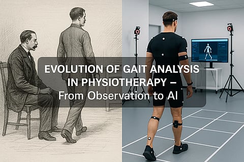
A split image that depicts earlier gait analysis by physician observation and other showing a modern gait lab.
What Are the Barriers to Access to Pediatric Gait Analysis?

Related Stories

No stories found.
logo
Medbound
www.medboundtimes.com