Researchers led by Queen Mary University of London have developed a non-invasive brush biopsy test that can help identify oral squamous cell carcinoma from suspicious oral lesions in under an hour, providing a quicker alternative to conventional surgical biopsies.
Published in the journal Biomarker Research, the study reports that the new diagnostic approach may help reduce the need for invasive scalpel biopsies. According to the researchers, the test could potentially prevent more than 90% of unnecessary biopsies among patients with low-risk oral lesions while enabling faster evaluation of suspicious mouth abnormalities.1
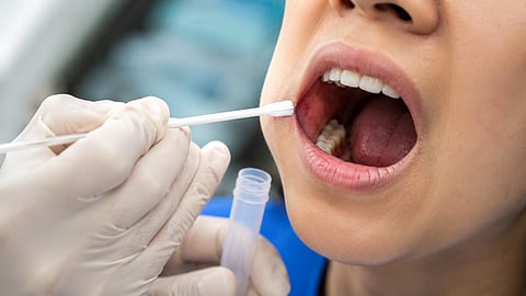
The diagnostic method, known as qMIDS V3 (quantitative Malignancy Index Diagnostic System), involves gently brushing the surface of a suspicious oral lesion to collect cells. Unlike a conventional biopsy, the procedure does not require cutting tissue.
The collected cells are analyzed using reverse transcription quantitative polymerase chain reaction (RT-qPCR) to measure the activity of four messenger RNA (mRNA) biomarkers, INHBA, S100A16, YAP1, and POLR2A, to determine whether cancer-associated molecular changes are present. Because this automated molecular assay bypasses the days of laboratory processing and slide staining required for traditional pathology, it can deliver results in under an hour.
The cross-continental study was a collaborative effort between the Queen Mary University of London and leading Indian institutions, including the All India Institute of Medical Sciences (AIIMS), King George's Medical University (KGMU), and Modern Dental College & Research Centre. To validate the test, researchers analyzed 1,090 oral brush samples collected from 545 adults between October 2021 and June 2024.
The cross-continental study was a collaborative effort between the Queen Mary University of London and leading Indian institutions, including the All India Institute of Medical Sciences (AIIMS), King George's Medical University (KGMU), and Modern Dental College & Research Centre. To validate the test, researchers analyzed 1,090 oral brush samples collected from 545 adults between October 2021 and June 2024. Each participant provided paired brush samples from the suspicious lesion and the contralateral clinically normal oral mucosa, resulting in two samples per participant.
The participants included individuals diagnosed with:
Oral squamous cell carcinoma
Oral leukoplakia
Oral lichen planus
Healthy oral mucosa (control group)
The researchers compared the brush test results with confirmed clinical diagnoses to evaluate the test's performance.
The study found that the four-gene qMIDS V3 test accurately distinguished oral cancer from common low-risk oral lesions.
The reported performance included:
95.7% sensitivity
95.1% specificity
95.5% overall diagnostic accuracy
Area under the curve (AUC): 0.975
These findings indicate that the test correctly identified most patients with oral cancer while also correctly excluding many patients without the disease.
According to The ASCO Post, lead researcher Muy-Teck Teh said,
We were genuinely astonished by the fact that the brush swab test performance is comparable to a microbiopsy. It suggests that the biological signal captured by these four genes is sufficiently strong and consistent that it can be detected even from the superficial exfoliated cells collected by a brush biopsy. The clinical implications are significant: patients no longer need even a minimally invasive procedure to benefit from molecularly guided triage.
Muy-Teck Teh, Lead Researcher, Professor of Molecular Oral Oncology, Queen Mary University of London.
Researchers estimate that more than 90% of patients with low-risk oral lesions could avoid unnecessary surgical biopsies if the brush test is used as an initial screening tool before tissue confirmation.
Many patients with suspicious mouth lesions undergo surgical biopsies that ultimately do not show cancer. According to the researchers, the new brush test could help identify patients who are at lower risk, potentially avoiding unnecessary invasive procedures.
The study estimated that more than 90% of patients with low-risk oral lesions could avoid surgical biopsy if the test is used as an initial screening tool before tissue confirmation.
Because the brush collects cells from a larger area of the lesion, it can also be repeated during follow-up visits with minimal discomfort.
Oral cancer survival is directly linked to how early it is found, yet our current diagnostic pathway is blunt—most patients with a suspicious lesion end up having an invasive biopsy even when the overwhelming likelihood is that it is benign. This test changes that. It gives clinicians a rapid, accurate, and noninvasive way to triage patients, and crucially, it can be repeated. That means we can now monitor patients with persistent premalignant lesions regularly and systematically—and pick up cancers much earlier than we would have been able to before.
Muy-Teck Teh, Lead Researcher, Professor of Molecular Oral Oncology, Queen Mary University of London.
A faster and less invasive diagnostic approach could support earlier evaluation of suspicious oral lesions, particularly in regions where oral cancer is common.
Oral cancer remains a significant public health challenge worldwide. According to data cited in the study, an estimated 422,000 new cases of lip and oral cavity cancer and approximately 229,000 deaths occurred globally in 2023. Asia accounts for more than two-thirds of newly diagnosed cases.
Early diagnosis allows patients to begin treatment sooner, which is associated with better clinical outcomes.
MedBound Times spoke with Dr. Bhuvan Nagpal, an oral and maxillofacial pathologist, clinical biochemist, and medico-legal consultant, to understand the importance of early detection of oral cancer.
Early detection of oral cancer is critical because it truly saves lives and preserves quality of life. The difference in outcomes is striking: when detected at an early stage, the 5-year survival rate is approximately 80–84%. In contrast, if the cancer is diagnosed late, after it has spread to lymph nodes or deeper tissues, survival drops dramatically to 30–40%.
Dr. Bhuvan Nagpal, MDS, Oral Pathology and Microbiology
Dr. Bhuvan Nagpal emphasized the importance of regular oral self-examinations and routine dental screenings for detecting oral cancer at an early stage.
An oral self-exam is basically when a person checks their own mouth regularly (say once a month) using a mirror and good lighting, looking for any of the “silent signs”.
Dr. Bhuvan Nagpal, MDS, Oral Pathology and Microbiology
He further elaborated,
Research from public health experts has concluded that conducting frequent cancer awareness and screening camps will detect oral cancers at an early stage in more people.
Dr. Bhuvan Nagpal, MDS, Oral Pathology and Microbiology
The current findings indicate that qMIDS V3 could serve as a rapid, non-invasive method for assessing suspicious oral lesions and helping clinicians decide which patients require confirmatory biopsy.
1. Teh, Min T., Rajashri Patil, Shubham A. Tekade, et al. "INHBA–S100A16 Dysregulation Enables a Non-Invasive Molecular Stratification Platform for Rapid Detection of Oral Squamous Cell Carcinoma: Results from a Large Diagnostic Case-Control Study." Biomarker Research 14, no. 76 (2026). https://doi.org/10.1186/s40364-026-00963-7.
(Rh/SS/MSM)