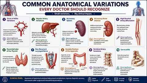
Every year, surgeons, radiologists, and clinicians encounter unexpected arteries, nerves, and organs that do not perfectly match textbook diagrams. Most of these differences are normal anatomical variations rather than abnormalities, but failing to recognize them can lead to excessive bleeding, nerve injury, incorrect diagnoses, prolonged operative time, or avoidable surgical complications. Understanding these common variations is therefore just as important as learning standard anatomy. Surgery, radiology, and many diagnostic and interventional procedures depend on a thorough knowledge of both normal anatomy and its commonly encountered variations.
Anatomical variations are naturally occurring structural differences that develop during embryological development and are usually asymptomatic. Most anatomical variations arise because blood vessels, nerves, and organs develop through complex embryological pathways. Small differences during fetal development can persist into adulthood without causing disease. Although often overlooked during anatomy education, these variations are frequently encountered in clinical practice and can significantly influence surgical planning, imaging interpretation, and patient outcomes.1,2
Failure to recognize an unexpected artery, nerve, or organ position may lead to excessive bleeding, nerve injury, prolonged operative time, misdiagnosis, or other preventable complications. Conversely, awareness of these variations allows clinicians to anticipate challenges, modify surgical approaches, and improve patient safety.1,3
Here are 10 clinically important anatomical variations that every medical student and healthcare professional should recognize to improve diagnostic accuracy, surgical safety, and patient outcomes.
Normal anatomy:
The Circle of Willis is an arterial ring at the base of the brain that connects the anterior and posterior cerebral circulations, providing collateral blood flow when a major cerebral artery is compromised.4
Common variation:
A complete, symmetrical Circle of Willis is present in only 20–25% of individuals. Common variations include hypoplasia or absence of the posterior communicating arteries, hypoplasia of the anterior communicating artery, and other variations that alter the pattern of collateral circulation.4
Clinical significance:
Variations of the Circle of Willis can reduce collateral blood flow during arterial occlusion, potentially influencing the severity of ischemic stroke. During an ischemic stroke, patients with an incomplete Circle of Willis may have reduced collateral circulation, potentially resulting in larger cerebral infarcts than those with a complete arterial ring. They are also important considerations during aneurysm surgery, carotid endarterectomy, and endovascular procedures, where vascular anatomy affects treatment planning.
Clinical Pearl:
A complete Circle of Willis is the exception rather than the rule. Recognizing these anatomical variations on CT or MR angiography helps guide stroke management and prevents misinterpretation of normal variants as vascular pathology.
Normal anatomy:
The common hepatic artery arises from the celiac trunk and continues as the proper hepatic artery, which divides into the right and left hepatic arteries.
Common variation:
Classic hepatic arterial anatomy is present in approximately 70% of individuals. The remaining 30% have variations, including replaced or accessory hepatic arteries. A replaced right hepatic artery arising from the superior mesenteric artery is one of the most common variants, occurring in 9–20% of individuals.1 These arterial patterns are commonly described using the Michels and Hiatt classification systems, which remain widely used in hepatobiliary surgery and liver transplantation planning.⁷˒⁸
Clinical significance:
Failure to recognize hepatic artery variations can increase the risk of hepatic ischemia, bile duct injury, and significant intraoperative bleeding during hepatobiliary, pancreatic, and liver transplant surgeries.
Clinical Pearl:
Preoperative CT angiography should be routinely reviewed before liver or pancreatic surgery to identify hepatic arterial variations and minimize surgical complications.
Normal anatomy:
The spleen is normally a single organ located in the left upper quadrant of the abdomen, receiving its blood supply from the splenic artery.
Common variation:
An accessory spleen (splenunculus) is present in approximately 10–30% of individuals. It is a congenital focus of normal splenic tissue, most commonly found near the splenic hilum or the tail of the pancreas.1,2 Unlike splenosis, which develops after splenic trauma or splenectomy through autotransplantation of splenic tissue, an accessory spleen is congenital and results from incomplete fusion of splenic tissue during embryological development.⁹˒¹⁰
Clinical significance:
Accessory spleens are usually asymptomatic but may be mistaken for enlarged lymph nodes, adrenal masses, or pancreatic tumors on imaging. During therapeutic splenectomy for conditions such as immune thrombocytopenia (ITP) or hereditary spherocytosis, failure to identify and remove an accessory spleen may result in persistent or recurrent disease.3
Clinical Pearl:
Always consider an accessory spleen when evaluating a left upper abdominal mass or persistent disease after splenectomy. Preoperative CT or MRI can help identify accessory splenic tissue.
Normal anatomy:
Each kidney is typically supplied by a single renal artery arising from the abdominal aorta, which enters the renal hilum before dividing into segmental branches.4
Common variation:
Accessory (supernumerary) renal arteries are present in 20–40% of individuals. These additional arteries may enter the kidney through the hilum or directly through the upper or lower pole, where they function as end arteries with little or no collateral circulation.4
Clinical significance:
Failure to recognize accessory renal arteries during nephrectomy, renal transplantation, or endovascular procedures may result in renal infarction, excessive bleeding, or postoperative complications. Multiple renal arteries also increase the technical complexity of kidney transplantation because each vessel may require separate vascular reconstruction. An accessory lower-pole renal artery may also compress the proximal ureter, contributing to ureteropelvic junction obstruction in some patients.¹¹
Clinical Pearl:
Preoperative CT angiography is essential before renal surgery or transplantation to identify accessory renal arteries and guide surgical planning.
Normal anatomy:
The brachial artery normally divides into the radial and ulnar arteries in the cubital fossa.6
Common variation:
In approximately 8% of individuals, the brachial artery divides higher in the arm than usual, resulting in a high brachial artery bifurcation.6
Clinical significance:
This variation may complicate arterial cannulation, catheterization, and vascular access procedures. A superficially located radial or ulnar artery may be mistaken for a vein, increasing the risk of accidental intra-arterial injection. High brachial artery bifurcation may also pose technical challenges during transradial catheterization and other upper-limb vascular interventions.⁶
Clinical Pearl:
Unexpected arterial anatomy should always be considered during upper-limb vascular procedures.
Normal anatomy:
The thyroid gland consists of two lateral lobes connected by an isthmus.1
Common variation:
A pyramidal lobe is present in approximately 40–50% of individuals and represents a persistent thyroglossal duct remnant.1
Clinical significance:
Residual thyroid tissue may remain after total thyroidectomy if the pyramidal lobe is overlooked.
Clinical Pearl:
Always inspect for a pyramidal lobe during thyroidectomy to reduce the risk of recurrent disease.
Normal anatomy:
The recurrent laryngeal nerve loops around the subclavian artery (right) or aortic arch (left) before ascending to the larynx.5
Common variation:
The non-recurrent laryngeal nerve (NRLN) is a rare anatomical variation of the recurrent laryngeal nerve (RLN) that results from abnormal embryological development. It is rare, occurring in approximately 0% to 4.76% of individuals, almost always on the right side.5
Clinical significance:
Failure to recognize this variation can result in permanent vocal cord paralysis during thyroid surgery.
Clinical Pearl:
An aberrant right subclavian artery on imaging should raise suspicion for a non-recurrent laryngeal nerve.
Normal anatomy:
The appendix arises from the posteromedial wall of the cecum.4
Common variation:
The retrocecal position is most common (60–65%), followed by the pelvic position (25–30%), while pre- and post-ileal positions are less frequent.4
Clinical significance:
The position of the appendix influences the presentation and diagnosis of acute appendicitis.
Clinical Pearl:
A retrocecal appendix may present with atypical flank or back pain rather than classic right iliac fossa tenderness.
Normal anatomy:
The vertebral arteries usually arise from the subclavian arteries and enter the transverse foramen of the sixth cervical vertebra.4
Common variation:
The left vertebral artery arises directly from the aortic arch in approximately 5% of individuals.4
Clinical significance:
Variations may complicate cervical spine surgery, angiography, and endovascular procedures. In particular, a left vertebral artery arising directly from the aortic arch may alter cerebral blood flow patterns and should be recognized before carotid or aortic arch interventions to reduce the risk of inadvertent vascular injury or procedural complications.⁴
Clinical Pearl:
Preoperative vascular imaging helps prevent inadvertent vertebral artery injury.
Normal anatomy:
Humans normally have 12 pairs of ribs, all of which arise from the thoracic vertebrae. A cervical rib is an additional rib that develops from the seventh cervical vertebra (C7).4
Common variation:
A cervical rib is present in approximately 0.5–1% of the general population and is more frequently observed in females. It may range from a small bony projection to a fully developed rib that articulates with or is connected by a fibrous band to the first rib.4
Clinical significance:
Although often asymptomatic, a cervical rib or its associated fibrous band may compress the lower trunk of the brachial plexus or the subclavian artery within the thoracic outlet. This can result in thoracic outlet syndrome (TOS), presenting with pain, numbness, paresthesia, or weakness of the upper limb. Less commonly, vascular compression may lead to subclavian artery stenosis, thrombosis, or distal embolization.
Clinical Pearl:
A cervical rib should be considered in patients with unexplained upper-limb neurological or vascular symptoms. A plain cervical spine radiograph is usually sufficient for diagnosis, while CT angiography or MRI can aid in surgical planning when intervention is required.
Human anatomy is inherently variable, and the "classic" textbook pattern represents only one of many normal anatomical configurations. Recognizing these variations is essential for accurate diagnosis, safe surgical practice, and appropriate interpretation of imaging studies.
Although anatomical variations are usually harmless, recognizing them is essential because normal does not always mean predictable. Whether interpreting CT scans, planning surgery, or examining patients, clinicians who understand anatomical diversity are better equipped to anticipate procedural challenges, reduce complications, and deliver safer patient care.
Are anatomical variations considered abnormalities?
No. Most anatomical variations are normal developmental differences that do not cause symptoms or impair function. However, recognizing them is important because they may influence imaging interpretation, surgical planning, and clinical procedures.
Why are anatomical variations important in surgery?
Unexpected anatomical variations can increase the risk of bleeding, nerve injury, organ damage, or technical difficulties during surgery and interventional procedures. Preoperative imaging and anatomical knowledge help surgeons anticipate these variations and improve patient safety.
What is the most common anatomical variation?
There is no single most common anatomical variation because prevalence differs by organ system. Examples include a pyramidal lobe of the thyroid (40 to 50%), accessory renal arteries (20 to 40%), accessory spleens (10 to 30%), and variations of the Circle of Willis, which are more common than the classic complete arterial configuration.
Can anatomical variations cause symptoms?
Most anatomical variations remain asymptomatic throughout life. However, some may become clinically significant by compressing nearby structures, altering blood flow, mimicking disease on imaging, or increasing the complexity of surgical and interventional procedures.
1. Sharma, Rashi, SPAS Nishan, Sunil Kumar Yadav, and Dharmendra Choudhary. 2025. "A Comprehensive Review of Anatomical Variations and Their Clinical Significance in Surgical Procedures." Journal of Ayurveda and Integrated Medical Sciences 10 (5): 136–146. doi:10.21760/jaims.10.5.20
2. Smith, Heather F. 2021. "Anatomical Variation and Clinical Diagnosis" Diagnostics 11, no. 2: 247. https://doi.org/10.3390/diagnostics11020247
3. Kowalczyk, Katarzyna, and Adrianna Majewski. 2021. "Analysis of Surgical Errors Associated with Anatomical Variations Clinically Relevant in General Surgery: Review of the Literature." Translational Research in Anatomy 23: 100107. https://doi.org/10.1016/j.tria.2020.100107.
4. Khan, Altamas, et al. 2025."Clinical Significance of Anatomical Variations in the Human Body: Implications for Surgeons and Radiologists." International Research Journal of Modernization in Engineering Technology and Science. https://doi.org/10.56726/IRJMETS93262.
5. Henry, Brandon M., Salvatore Sanna, Michael J. Graves, Jacek Vikse, Barbara Sanna, Iwona M. Tomaszewska, R. Shane Tubbs, Jan A. Walocha, and Krzysztof A. Tomaszewski. 2017. "The Non-Recurrent Laryngeal Nerve: A Meta-Analysis and Clinical Considerations." PeerJ 5: e3012. https://doi.org/10.7717/peerj.3012.
6. Rodríguez-Niedenführ, M., T. Vázquez, L. Nearn, B. Ferreira, I. Parkin, and J. R. Sañudo. 2001. "Variations of the Arterial Pattern in the Upper Limb Revisited: A Morphological and Statistical Study, with a Review of the Literature." Journal of Anatomy 199 (5): 547–566. https://doi.org/10.1046/j.1469-7580.2001.19950547.x.
7. Michels, Nicholas A. 1966. “Newer Anatomy of the Liver and Its Variant Blood Supply and Collateral Circulation.” The American Journal of Surgery 112 (3): 337–347. https://doi.org/10.1016/0002-9610(66)90201-7.
8. Hiatt, Jonathan R., Joubin Gabbay, and Ronald W. Busuttil. 1994. “Surgical Anatomy of the Hepatic Arteries in 1000 Cases.” Annals of Surgery 220 (1): 50–52. https://doi.org/10.1097/00000658-199407000-00008.
9. Moore, Keith L., T. V. N. Persaud, and Mark G. Torchia. 2023. The Developing Human: Clinically Oriented Embryology. 12th ed. Philadelphia: Elsevier. https://www.educate.elsevier.com/book/details/9780443116988.
10. Mortelé, Koenraad J., Bieke Mortelé, and Stuart G. Silverman. 2004. “CT Features of the Accessory Spleen.” American Journal of Roentgenology 183 (6): 1653–1657. https://doi.org/10.2214/ajr.183.6.01831653
11. Sampaio, Francisco J. B., and M. A. R. F. Passos. 1992. “Renal Arteries: Anatomic Study for Surgical and Radiological Practice.” Surgical and Radiologic Anatomy 14 (2): 113–117.