- Home
- About Us
- Contact Us
- Biography
- Column
- MedBound Hub
- InternshipInternship
- Daily Pulse
- Medicine
- Physical Therapy
- InterviewInterview
- Pharmacy
- Biotechnology
- College/InstituteCollege/Institute
- Dentistry
- Blog
- Wellness & Nutrition
- Nursing
- Opinion

A 30-year-old woman from coastal New England was diagnosed with a parasitic infection affecting the brain after returning from a three-week trip to Thailand, Japan, and Hawaii, according to a case published in the New England Journal of Medicine.1 Her symptoms began about eight days before hospital admission and progressively worsened over several days.
The patient initially reported fatigue, which she attributed to jet lag. She then developed a burning sensation in her feet that spread to her legs, trunk, and arms and worsened with light touch, Dr. Carlos A. Portales Castillo, who later managed her care at Massachusetts General Hospital, reported.
She visited two emergency departments, where examinations were reported as normal. Blood tests showed elevated eosinophil levels, but no diagnosis was established. She was discharged with instructions for follow-up. As her condition progressed, she developed a headache that did not respond to medication and recorded a fever at home.
The patient’s condition worsened when she developed confusion. According to People, she became disoriented and was unable to recognize her situation, prompting her roommate to seek further medical care.
Her roommate brought her to Massachusetts General Hospital after she woke up confused, believing she needed to prepare for a trip and was unable to be redirected. The confusion persisted for several hours, as described by Dr. Carlos A. Portales Castillo.
On admission, clinicians noted altered mental status. A lumbar puncture revealed elevated cerebrospinal fluid pressure and a high white-cell count, including eosinophils. These findings confirmed eosinophilic meningitis, a condition often linked to parasitic infections.
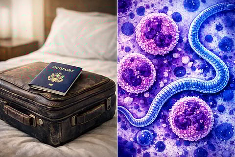
According to the journal, Dr. Joseph Zunt, a neurologist and infectious disease specialist who evaluated the case, considered eosinophilic meningitis due to Angiostrongylus cantonensis after reviewing the patient’s travel history and activities during her trip.
She swam in the ocean several times and frequently ate both salad and sushi.
Dr. Joseph Zunt, Neurologist and Infectious disease specialist
The patient reported consuming sushi and salads and swimming in the ocean during her stay in Hawaii. These exposures were considered relevant, as infection can occur through ingestion of contaminated food or contact with environments where infected snails or slugs are present.
As Dr. Zunt noted, the rat lungworm parasite is commonly found in Hawaii and can be readily transmitted.
Infection can be acquired through multiple sources: ingestion of raw or undercooked infected snails or slugs; ingestion of vegetables or fruits contaminated by infected snails, slugs, or flatworms or by slime from snails or slugs that contains infectious larvae; or ingestion of infected paratenic hosts (e.g., land crabs, freshwater prawns, or frogs) that have consumed an infected snail.
Dr. Joseph Zunt, Neurologist and Infectious disease specialist
Humans become infected when larvae enter the body, often through contaminated vegetables or undercooked intermediate hosts. The larvae can migrate to the central nervous system, where they cause inflammation.
Caused by the parasitic roundworm Angiostrongylus cantonensis
Rats act as the primary hosts; snails and slugs serve as intermediate hosts
Humans are accidental hosts who become infected after ingesting larvae
Can lead to eosinophilic meningitis, an inflammation of the brain and spinal cord coverings
Common in tropical and subtropical regions
Endemic in Southeast Asia and Pacific regions, including Hawaii
Increasingly reported in other parts of the world due to expanding distribution
Infection occurs through ingestion of parasite larvae
Consumption of raw or undercooked snails or slugs
Eating vegetables or fruits contaminated by snails, slugs, or their slime
Consumption of infected intermediate or paratenic hosts such as freshwater prawns, crabs, or frogs
Larvae enter the body after ingestion
Migrate through the bloodstream or peripheral nerves
Reach the central nervous system, including the brain
Trigger inflammation leading to eosinophilic meningitis
Incubation period typically ranges from 1 to 2 weeks
Headache
Burning or tingling sensations (dysesthesia, paresthesia)
Hypersensitivity to touch (allodynia)
Fever in some cases
Confusion or altered mental status in advanced stages
Based on symptoms and recent travel history
Blood tests may show eosinophilia
CSF analysis confirms eosinophilic meningitis (raised cells, eosinophils)
NAAT of CSF detects A. cantonensis DNA
Corticosteroids to reduce inflammation
Albendazole as antiparasitic therapy
Supportive care for symptoms
Most patients improve with treatment
The patient received corticosteroids to reduce inflammation and albendazole as antiparasitic therapy. Her symptoms improved during hospitalization, and she was discharged after six days.
This case demonstrates how travel history, dietary exposure, and laboratory findings such as eosinophilia contribute to identifying parasitic infections. Early use of cerebrospinal fluid analysis and molecular testing supports accurate diagnosis and timely treatment.
1. Zunt, Joseph R., Amy K. Barczak, and Daniel Y. Chang. “Case 5-2025: A 30-Year-Old Woman with Headache and Dysesthesia.” New England Journal of Medicine 392, no. 7 (2025): 699–709. https://doi.org/10.1056/NEJMcpc2412514.
(Rh/SS)